
About Myopic Choroidal Neovascularization (mCNV)
What causes mCNV?
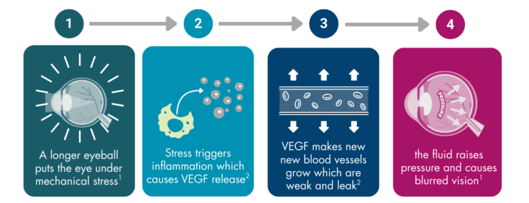
The vision loss experienced in mCNV is caused by the growth of abnormal blood vessels in the macula. These blood vessels grow due to structural changes in the eye associated with near-sightedness.1
Near-sighted individuals have a longer eyeball than the average person; it is stretched from front to back. As a result, the retina at the back of the eye, responsible for vision, can become thin and prone to changes such as the growth of blood vessels through the release of inflammatory chemicals like vascular endothelial growth factor (VEGF).1,2 These newly formed blood vessels are weak and can leak, leading to the build-up of fluid in the back of the eye, known as macular oedema.
References
- NHS Manchester Royal Eye Hospital Retinal Services Information for Patients. Treatment of Myopic Choroidal Neovascularisation (CNV). Available at: https://mft.nhs.uk/app/uploads/sites/2/2018/04/REH-213.pdf. Last accessed May 2025.
- Kumar A et al. Indian J Ophthalmol. 2017;65(2):85.